+971 4 395 55 91
info@dlsclinic.com
Villa 467B Beach Road,
Jumeirah 3, Dubai, UAE

Obstetrics

Pregnancy is one of the most remarkable milestones in every woman’s life.

Dr. Leila Soudah makes you and your baby feel safe in every step of the pregnancy, childbirth and after delivery.

The ability of a woman to conceive and give birth to a baby is a wonderful blessing that brings joy to a couple. It builds a strong foundation for a family.

The clinic offers the following services suitable for the different stages of pregnancy you are in:

ULTRASOUND

It is a fascinating experience for a woman to see what her baby looks like while inside the womb. The ultrasound machines shows images of how well the baby is developing inside the uterus. It displays the size of the baby, its position in the womb, the heartbeat, internal organs, the amount of amniotic fluid, the expected date of delivery and will also show any abnormalities if present. The ultrasound is not the only for taking beautiful pictures of the baby but also to detect abnormalities as early as possible.

The clinic uses Voluson E10 B18 GE ultrasound system, which is an ADVANCED HIGH DEFINITION LIVE ULTRASOUNDS.

The machine has the following specifications1:
  • HDLive: An extraordinary rendering method generating amazingly realistic images of the human fetus to help improve diagnostic confidence.
  • Advanced Probe Technology: View images created with advanced Voluson probes that demonstrate extraordinary image quality.
  • Early Health: View fine details that help clinicians see early into the first trimester.
  • Fetal Cardiac Assessment: Voluson advanced fetal heart tools help enhance visualization of the fetal heart anatomy.
  • Fetal CNS: Voluson advanced imaging tools enable detailed assessment of the fetal central nervous system
  • Image Quality: Experience extraordinary vision created by Voluson E8 ultrasound systems
  • Silhouette: Emphasizes the contour surface structures and improve the impression even more.

FIRST TRIMESTER SCREENING

It is a painless, in-clinic ultrasound and blood procedure, an easy screening test that a pregnant woman can do and go back to her normal routine after the procedure. This test will rule out birth defects like neural tube, chromosomal abnormality such as trisomy 13, 18, 21, as well as the risk for pregnancy complications detailing the following:
  • Risk for fetal growth restriction before 37 weeks
  • Risk for pre-eclampsia before 34 weeks
  • Risk for pre-eclampsia before 37 weeks
  • Risk for pre-eclampsia before 42 weeks

Every pregnant patient should be offered this kind of test regardless of her age. This screening consists of TWO PARTS:

  1. BLOOD TEST
    This is done ideally during 11 to 12 weeks of pregnancy. This is a screening test for an early detection of aneuploidy pregnancy. This screening is done by the combination of two biochemical markers which are the free Beta HCG and Pregnancy Associated Plasma Protein (PAPP-A). The patient is requested to set an appointment early in the morning since blood samples are referred to an outsource laboratory in Germany.
    Be reminded that the level of these results are correlated with the NT scan to calculate the risk of aneuploidy
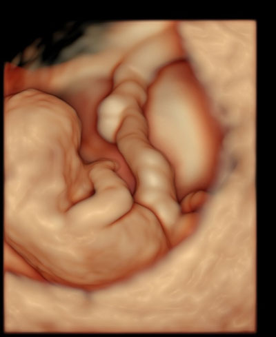
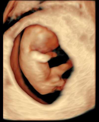
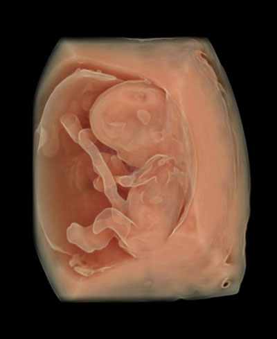
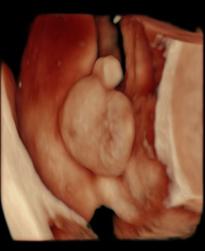
  2. NT SCAN/ULTRASOUND
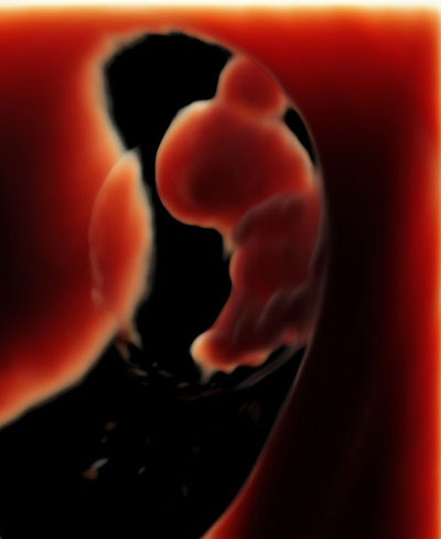
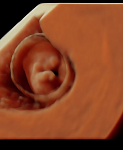
    This is ideally performed from the start of 11 weeks to 14 weeks of pregnancy.
    Dr. Leila Soudah has been a licensed sonographer and has a certificate of competence in performing the first trimester screening under both the English and German Fetal Medicine Foundation since 2004. This enables her to perform this test and to calculate the risk of abnormality for your baby. Dr. Leila is listed as a sonographer with good audit results under FMF England.

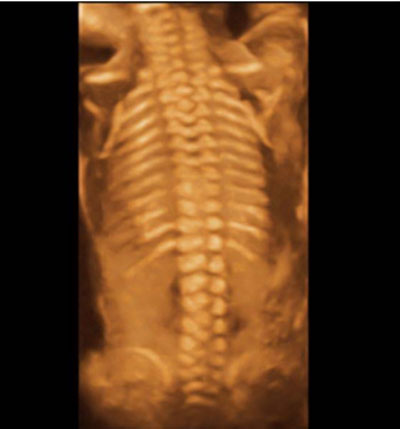
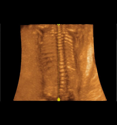
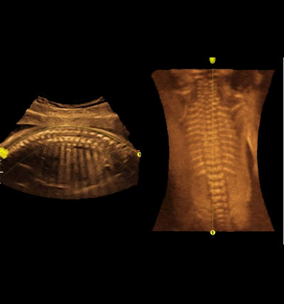
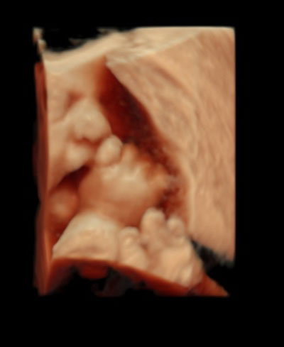
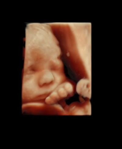
The Scan/Ultrasound demonstrates the following FIRST TRIMESTER MARKER:
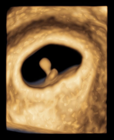
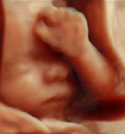
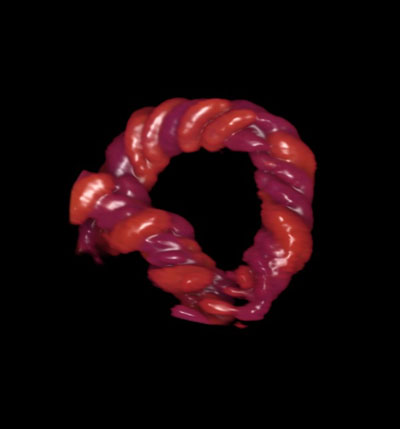
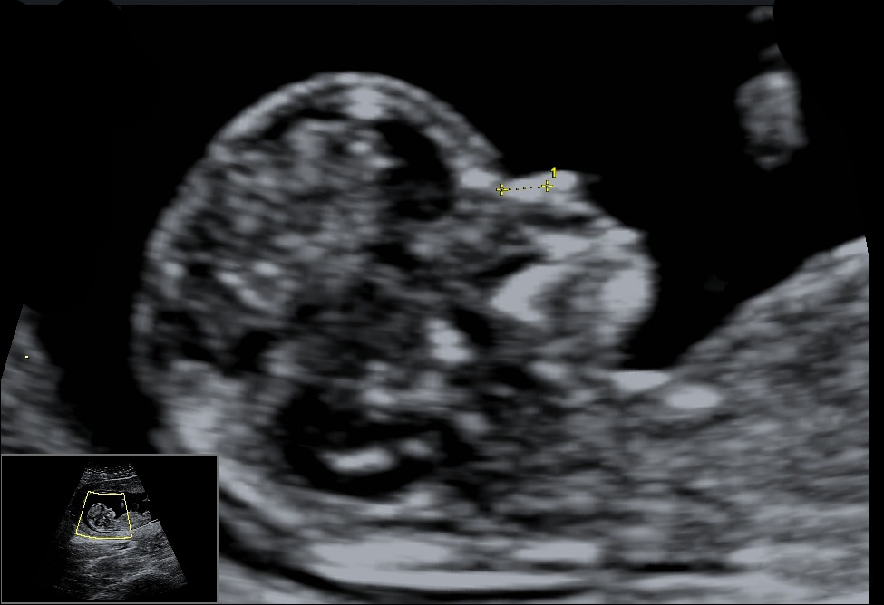

Nuchal Translucency
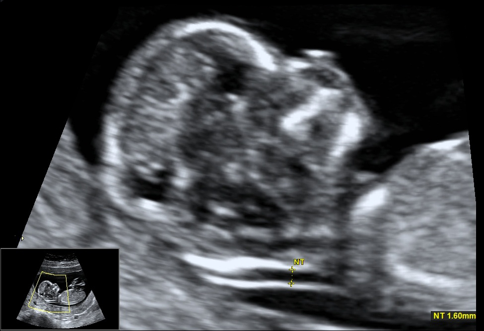
A nuchal translucency ultrasound measures the collection of fluid under the skin behind the fetal neck. NT thickness could increase for a fetus with chromosomal abnormalities, cardiac defects and other genetic syndromes. In correlation with the maternal serum free Beta HCG and PAPP-A provides an effective method for early screening for trisomy 13, 18, 21. The combination of Nuchal translucency and maternal serum free Beta HCG and PAPP-A increases detection up to 90%. There is now evidence that the detection rate can increase to about 95% and the false positive rate can be reduced to 2.5 % by also examining the following markers:
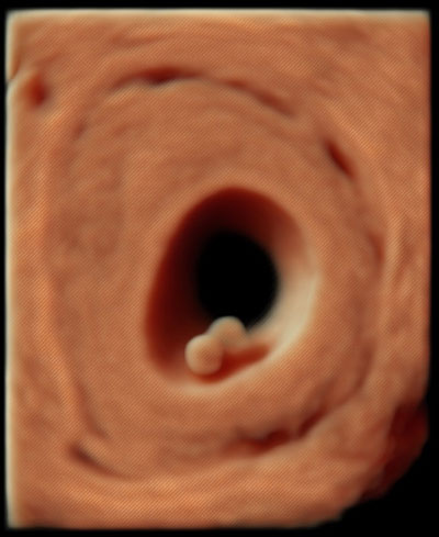
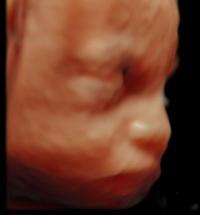
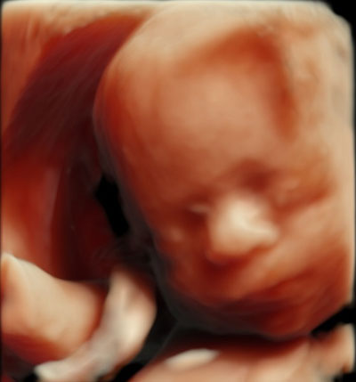
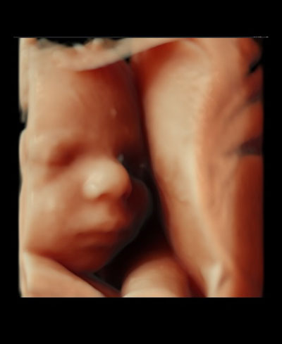
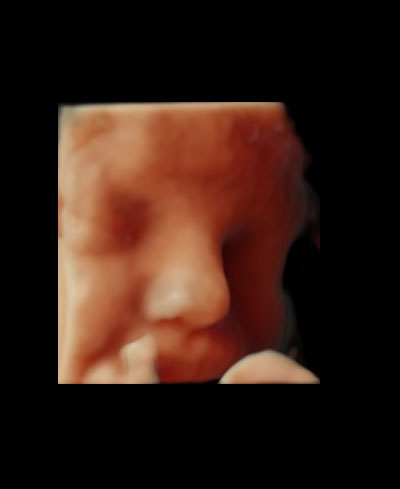
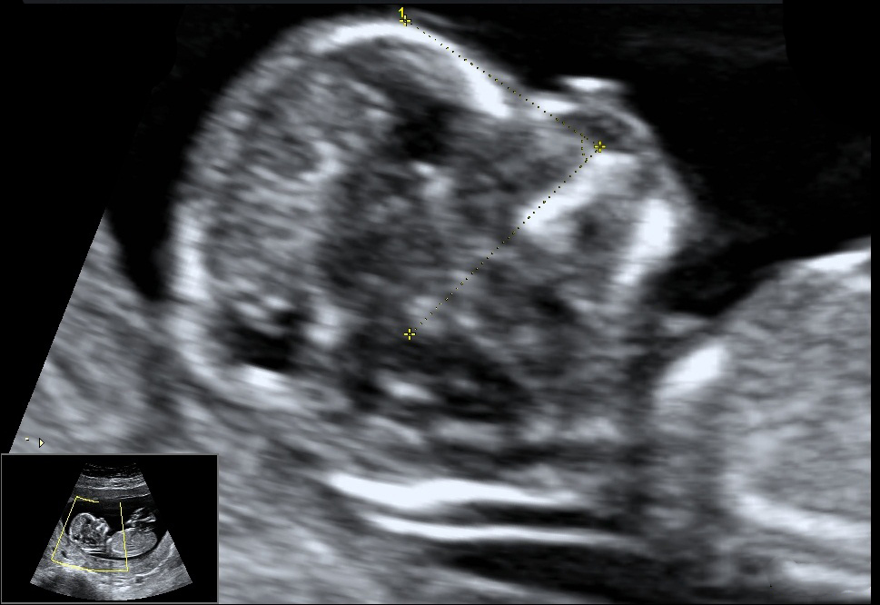
  • Nasal Bone: These are two oblique bones, placed side by side at the middle and upper part of the face and form by a junction in "the bridge" of the nose. For fetuses with trisomy 21 and other chromosomal abnormalities, the nasal bone is hypo plastic or not visible at 11-13 weeks of gestation.
  • Facial Angle: It is the wide angle in a line drawn over the palate and between the maxilla and the forehead. Recent studies suggest that fetuses with trisomy 21 have a flat profile because the maxilla (upper jaw) is small and set back.
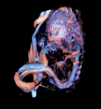
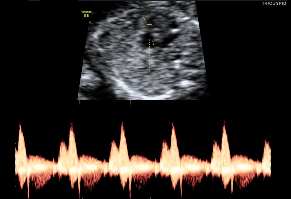
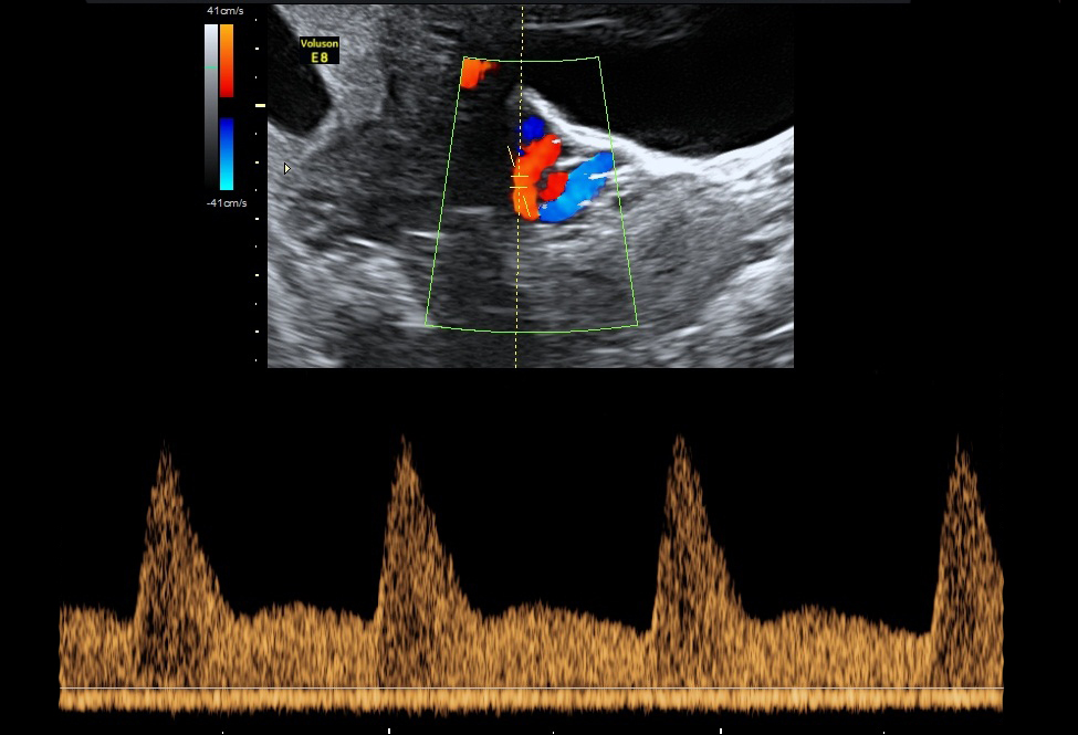
  • Tricuspid Valve: Flow of blood between right atrium to right ventricle. This is one of the markers that contribute in the screening for both major cardiac defects.
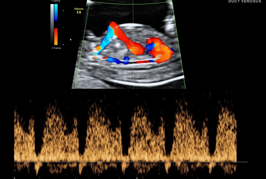
  • Ductus Venosus: Blood flow of the umbilical vein directly to the fetal heart. It is part of the fetal circulation. Presence of normal flow will reduce the risk for trisomy 21 and cardiac abnormalities and the presence of reversed A- wave will increase the risk.
  • Uterine Artery PI: It is the two arteries both left and right that supplies blood to the uterus in females. By using maternal history, as well as the uterine artery blood flow and the blood pressure from both arms, we can calculate the risk for complications that may arise during the pregnancy. Some of these complications include pre-eclampsia and hypertension.


Harmony Test
The Harmony Test is a non-invasive method for the detection of fetal trisomies in an unborn child, based on the analysis of genetic material (fetal DNA) that circulates in the mothers' blood. It is an early and reliable prenatal screening test for the most common chromosomal disorders. It can be performed using the mother’s blood after completion of the 10th week of gestation.
  1. TRISOMY 21
    Trisomy 21 is due to an extra chromosome 21 and is the most common trisomy at the time of birth. Trisomy 21, also known as Down syndrome, is associated with mild to moderate intellectual disabilities, which may also lead to digestive disease and congenital heart defects.
  2. TRISOMY 18
    Trisomy 18 is due to an extra chromosome 18. Trisomy 18, also called Edwards syndrome, is associated with a high rate of miscarriage. Infants born with trisomy 18 often have congenital heart defects as well as various other medical conditions, shortening their lifespan.
  3. TRISOMY 13
    Trisomy 13 is due to an extra chromosome 13. Trisomy 13, also called Patau syndrome, which is associated with a high rate of miscarriage. Infants born with trisomy 13 usually have severe congenital heart defects and other medical conditions. Survival beyond the first year is rare.

Highest Detection Rate
99.7% detection rate for Trisomy 21 in published studies

Lowest False Positive Rate
only 0.06% for Trisomy 21
Women above a certain age have a higher false positive rate for sex chromosome disorders. The Harmony Test false positive rate for trisomy 21 is 0.06%, and is slightly higher for sex chromosome disorders.

High Success Rate
Characterized by a high success rate. According to their own data, 98% of the tests were successful at the first attempt.

Application of the Harmony Test
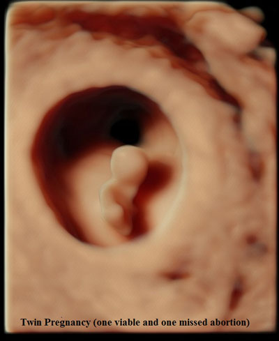
The test can be applied for all singleton or twin pregnancies, even if the pregnancy is the result of an in-vitro fertilization including egg donation.
In the case of twins, a determination of sex chromosomes disorders (X,Y) and fetal sex is not possible. (REFERENCE: CENATA GmBH)


Amniocentesis
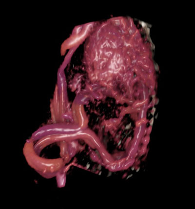
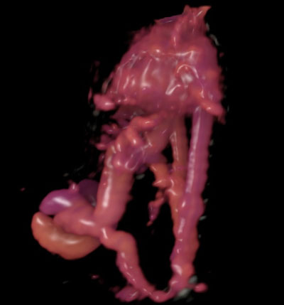
It is an invasive medical procedure used in prenatal diagnosis for chromosomal abnormalities and fetal infections, in which a small amount of amniotic fluid that contains the fetal tissue is aspirated in the amniotic sac surrounding the fetus under the guide of the ultrasound. This procedure is ideally done during weeks 15 to 20 of pregnancy.

SECOND TRIMESTER SCREENING

During this stage, typically you will see the Doctor once every four weeks unless you have a condition or complication that will call for more frequent visits for checkups. Here, blood tests will be done, such as short glucose tolerance test to rule out gestational diabetes. In addition, when a first trimester screening shows high risk, a fully integrated screening is offered. Second trimester screenings include the following:

  • Anomaly or detailed scan:
    This is a one-hour ultrasound procedure where Dr. Leila Soudah takes a precise and closer look to examine each part of the fetal body, determining the position of the placenta, assessing the amount of amniotic fluid, and measuring fetal growth. Special attention is paid to the vital developing organs such as the brain, face, spine, heart, stomach, bowel, kidneys and limbs.
    At this point, through the high definition ultrasound imaging technology, Dr. Leila, a licensed and experienced sonographer; will be able to pinpoint multiple potential problems that can arise in developing fetuses. Although not all possible physical conditions can be picked up by the scan, a significant proportion of major irregularities can be found and the necessary precautions will be taken and the right care will be advised along the pregnancy.
    While undergoing this procedure, the Doctor will tell you about everything that she sees, unless you advise that there are certain things that you don't want to know, such as the baby's gender.
    Furthermore, if serious problems are visible during the scan, Dr. Leila Soudah will give and support the opportunity to discuss all possible options, appropriate medical attention and suitable decisions.

  • Fetal echocardiography:
    It is an ultrasound that requires special doctor skills to examine the heart shape, size, position, blood flow and function. This information is essential in order for the doctor to determine things such as the mode of delivery and which hospital will be suitable for the right neo-natal care for your baby.

  • Fetal Fibronectin:
    Fetal Fibronectin in a cervicovaginal secretion used as an aid to rapidly assess the risk of preterm delivery. This test is done from the beginning of 28 weeks of pregnancy onwards. This is a painless procedure where Dr. Leila collects a swab from the pregnant patient’s cervix via a special transport swab. This test is more accurate in combination with the cervix length assessment (measuring the cervix length). In the case of a positive result, an increased risk for preterm delivery is highly likely. The doctor will provide you with the appropriate advice and medication to prevent incidence of preterm labor and subsequent delivery.

THIRD TRIMESTER SCREENING

The third trimester is the best time for evaluating fetal growth, the quantity of amniotic fluid, as well as the vessels of the mother and the baby, thus recognizing in time if the placenta functions properly or if there are any dangers for the baby. This is performed especially in cases of abnormal second trimester screening tests, multiple pregnancies, low level of amniotic fluid and risk of insufficient supply to the placenta.

  • Group B Streptococcus Screening:
    Vaginal and rectal swabs are taken at 35 to 37 weeks of pregnancy to detect group B strep bacteria. Although group B strep can be present in up to 30% of all healthy women, it's the leading cause of life-threatening infections in newborns, as well as causing mental retardation, impaired vision, and hearing loss. Women who test positive are treated in pattern presented with the lab results during delivery to protect the baby from acquiring the infection at birth.

  • Doppler Ultrasound/ Doppler Study:
    A procedure used to check the blood flow through the fetus's umbilical cord.
    Here at Dr. Leila Soudah Clinic, Color Doppler is used as a standard ultrasound method to produce a picture of a blood vessel. Also, a computer converts the Doppler sounds into colors that are overlaid on the image of the blood vessel representing the speed and direction of blood flow through the vessel. Power Doppler is a special type of color Doppler. This kind of test is compulsory to pregnant patients with a history of previous abortions, small babies or premature deliveries. Doppler Ultrasounds give you information about the baby’s blood supply.

  • Cardiotocography:
    This is done starting from 28 weeks of pregnancy for assessing fetal well-being, monitoring and recording the fetal heartbeat, and uterine contractions to prevent adverse fetal outcome such as pre-mature delivery.

  • Fetal Growth Scan:
    The purpose of this scan is to check that the baby is growing well and that the pregnancy is developing normally. Furthermore, this scan is particularly important if you have had pregnancy complications or problems in a previous pregnancy.
    The growth of the baby will be assessed by measuring the head, abdomen and femur length (thigh bone). This way the doctor can estimate the baby's weight and give you an idea as to how big it will be at birth. The appearance and position of the placenta is also checked, and the amniotic fluid around the baby is measured.
    This scan is significant for you as well, as it gives you an idea as to what the mode of delivery may be. This enables you to prepare yourself for the coming of the baby.
1Source: GE Healthcare